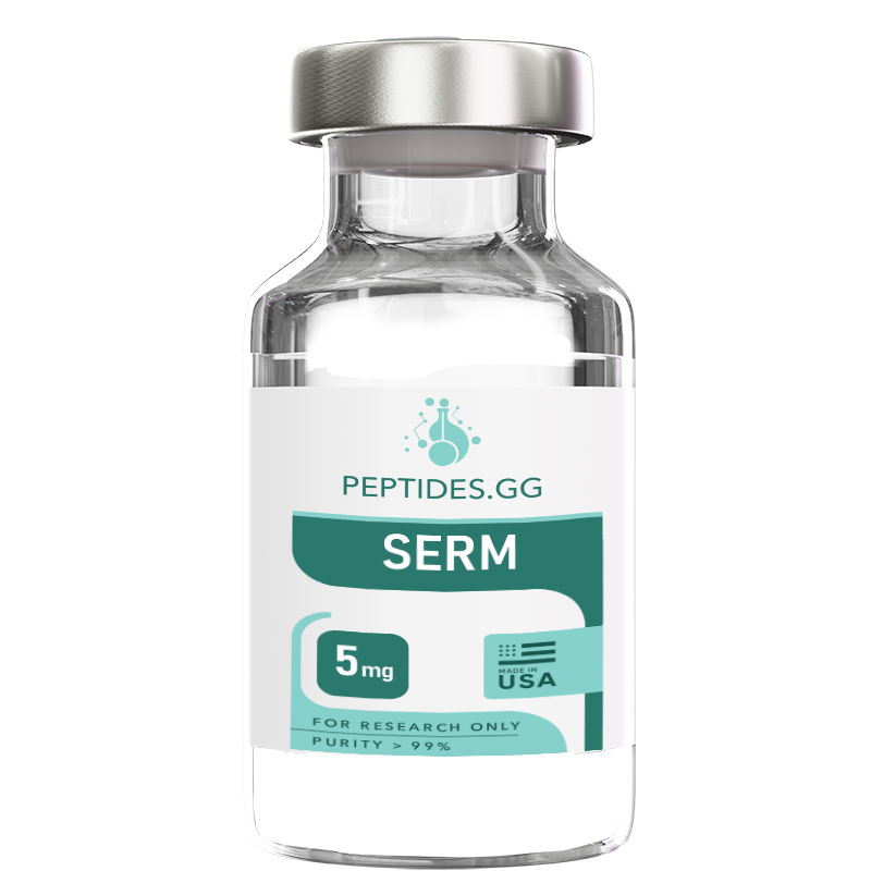
Buy SERM peptide for research applications. High-purity SERM research peptide available for laboratory studies and scientific investigation. Shop premium quality research-grade peptides manufactured in the USA with comprehensive Certificate of Analysis documentation.
Important: All products are intended as research chemicals only for laboratory and in vitro testing and experimentation. All product information is educational and not to be taken as medical advice. No products are for human or animal use.
SERM
$25.00 – $70.00
- Free Delivery on all orders over $200
- Earn 5% Store Credit with Every Order
- Same Day Shipping Before 1 PM PST
- 10% Discount for Cryptocurrency Payments
14-day money-back guarantee
If you are not satisfied with the product, simply return it and we will refund your money

Buy SERM peptide for research applications. High-purity SERM research peptide available for laboratory studies and scientific investigation. Shop premium quality research-grade peptides manufactured in the USA with comprehensive Certificate of Analysis documentation.
Important: All products are intended as research chemicals only for laboratory and in vitro testing and experimentation. All product information is educational and not to be taken as medical advice. No products are for human or animal use.
Research Overview
SERMs serve as critical research tools for investigating estrogen receptor biology and tissue-selective hormone signaling mechanisms in laboratory settings. These compounds revolutionized understanding of nuclear receptor pharmacology by demonstrating that receptor ligands could produce tissue-specific agonist or antagonist effects rather than uniform systemic responses. Research applications span endocrinology, oncology, bone biology, cardiovascular research, and reproductive biology, making SERMs among the most extensively studied nuclear receptor modulators.
The SERM concept emerged from clinical observations that compounds structurally related to estrogen could block estrogen effects in breast tissue while mimicking estrogen effects in bone and lipid metabolism. This tissue selectivity arises from multiple mechanisms including differential estrogen receptor subtype expression (ERα vs. ERβ), tissue-specific co-regulator expression patterns, receptor conformation changes induced by different ligands, and promoter-specific effects on gene transcription.
Laboratory studies investigate SERMs’ effects on estrogen receptor binding kinetics, receptor conformation dynamics, co-activator and co-repressor recruitment, gene expression patterns, and downstream cellular responses. Research protocols examine these effects in cell culture systems expressing different estrogen receptor subtypes, tissue-specific experimental models, and molecular studies of receptor-ligand-coregulator complexes. The wealth of SERM research has established fundamental principles of nuclear receptor pharmacology applicable to other receptor systems including androgen receptors, progesterone receptors, and metabolic nuclear receptors.
Molecular Characteristics and SERM Classes
Tamoxifen (First-Generation SERM):
- CAS Number: 10540-29-1 (base), 54965-24-1 (citrate salt)
- Molecular Weight: 371.51 Da (base), 563.64 Da (citrate)
- Molecular Formula: C₂₆H₂₉NO (base)
- Structure: Triphenylethylene derivative with dimethylaminoethoxy side chain
- ER Binding: Non-selective ERα/ERβ antagonist in breast, partial agonist in bone/uterus
- Metabolism: Extensive hepatic metabolism to active metabolites (4-hydroxytamoxifen, endoxifen)
- Research Applications: Breast cancer models, bone metabolism studies, lipid metabolism research
Tamoxifen represents the prototypical SERM with over five decades of research establishing tissue-selective mechanisms. The compound exists as geometric (E/Z) isomers, with trans-tamoxifen showing predominant biological activity. Tamoxifen’s triphenylethylene structure positions the basic dimethylaminoethoxy side chain to interfere with helix 12 positioning on the estrogen receptor, preventing full agonist conformation.
Raloxifene (Second-Generation SERM):
- CAS Number: 84449-90-1 (base), 82640-04-8 (hydrochloride)
- Molecular Weight: 473.58 Da (base), 510.05 Da (HCl)
- Molecular Formula: C₂₈H₂₇NO₄S
- Structure: Benzothiophene derivative with piperidine side chain
- ER Binding: ERα-preferential binding, pure antagonist in breast and uterus
- Advantages: Reduced uterine stimulation compared to tamoxifen
- Research Applications: Bone formation studies, breast cancer prevention models, cardiovascular research
Raloxifene’s benzothiophene core structure provides improved tissue selectivity profile compared to tamoxifen, particularly reduced uterine stimulation. The piperidine basic side chain interacts with the estrogen receptor to block co-activator recruitment more effectively in uterine tissue than tamoxifen.
Toremifene (Tamoxifen Analog):
- CAS Number: 89778-26-7 (base), 89778-27-8 (citrate)
- Molecular Weight: 405.96 Da (base)
- Molecular Formula: C₂₆H₂₈ClNO
- Structure: Chlorinated triphenylethylene (4-chloro derivative of tamoxifen)
- ER Binding: Similar to tamoxifen with subtle metabolic differences
- Research Applications: Comparative metabolism studies, tamoxifen alternative research
Toremifene differs from tamoxifen by a single chlorine atom on the ethyl group, resulting in altered metabolic profile. This structural similarity makes toremifene valuable for investigating how minor structural modifications affect SERM pharmacology and metabolism.
Clomiphene (Mixed Isomer SERM):
- CAS Number: 911-45-5 (base), 50-41-9 (citrate)
- Molecular Weight: 405.96 Da
- Molecular Formula: C₂₆H₂₈ClNO
- Structure: Triphenylethylene with chlorine substitution, exists as E/Z isomer mixture
- ER Binding: Hypothalamic-pituitary axis antagonism leading to gonadotropin release
- Isomers: Zuclomiphene (cis, more estrogenic) and enclomiphene (trans, more antiestrogenic)
- Research Applications: Reproductive axis studies, ovulation induction mechanisms, gonadotropin regulation
Clomiphene is typically used as a mixture of geometric isomers with distinct pharmacological properties, providing research tools for studying isomer-specific effects on estrogen receptor signaling.
Mechanisms of Tissue Selectivity
SERM tissue selectivity arises through multiple interconnected molecular mechanisms that represent fundamental principles of nuclear receptor pharmacology:
Receptor Conformation and Structure:
Crystallographic studies reveal that estrogen binding positions helix 12 of the ligand-binding domain to create a hydrophobic groove accommodating the LXXLL motif of co-activator proteins. SERM binding displaces helix 12 to an alternative position that blocks this co-activator binding surface. Different SERM structures induce subtly different helix 12 positions, explaining varying degrees of agonist vs. antagonist activity across SERMs and tissues.
The basic side chain present in most SERMs (dimethylaminoethoxy in tamoxifen, piperidine in raloxifene) extends toward helix 12 and physically prevents it from adopting the full agonist position. The size, flexibility, and chemical properties of this side chain influence the degree of helix 12 displacement and subsequent co-regulator recruitment preferences.
Co-regulator Expression Patterns:
Tissues express different repertoires of co-activators (SRC-1, SRC-2/TIF2/GRIP1, SRC-3/AIB1/ACTR) and co-repressors (NCoR, SMRT). In tissues with high co-activator to co-repressor ratios, SERM-receptor complexes may recruit sufficient co-activators to produce partial agonist effects. In tissues with lower co-activator availability or higher co-repressor levels, the same SERM-receptor complexes preferentially recruit co-repressors, producing antagonist effects.
Research demonstrates that experimental manipulation of co-regulator expression levels can convert a SERM from antagonist to agonist (by overexpressing co-activators) or enhance antagonist activity (by reducing co-activator availability). This demonstrates the critical importance of cellular context in SERM pharmacology.
Estrogen Receptor Subtype Distribution:
ERα and ERβ show distinct tissue expression patterns and some functional differences in response to ligands. ERα predominates in uterus, breast, and bone, while ERβ shows higher expression in ovary, prostate, and certain cardiovascular and central nervous system tissues. Some SERMs demonstrate preferential binding to one receptor subtype.
Research using ERα and ERβ knockout mice or cells transfected with individual receptor subtypes demonstrates that tissue-specific receptor subtype expression contributes to SERM tissue selectivity. However, most tissue selectivity arises from factors beyond simple receptor subtype distribution.
Promoter Context and ERE Architecture:
Estrogen-responsive genes contain various estrogen response element (ERE) sequences and AP-1 sites in their promoters. Classical palindromic EREs (GGTCAnnnTGACC) support strong estrogen agonist activity, while imperfect EREs or AP-1 sites show different responses to SERMs. SERM-receptor complexes demonstrate different transcriptional activities on classical EREs vs. AP-1 sites, contributing to gene-specific and tissue-specific effects.
Some genes show SERM agonist activity while others show antagonist activity within the same cell type, explained by promoter architecture differences. This gene-selective effect adds another layer to SERM pharmacology beyond tissue selectivity.
Research Applications
Estrogen Receptor Biology Research
SERMs provide essential tools for dissecting estrogen receptor structure-function relationships and receptor pharmacology principles applicable across nuclear receptor superfamily:
Receptor Binding and Affinity Studies:
- Competition binding assays using [³H]-estradiol or fluorescent estradiol analogs
- Determination of binding affinity (Ki or IC₅₀) for ERα and ERβ
- Kinetic analysis using surface plasmon resonance or biolayer interferometry
- Structure-activity relationship studies comparing SERM analogs
- Radioligand binding to membrane vs. nuclear estrogen receptors
Research protocols typically employ purified recombinant estrogen receptor ligand-binding domains or cell lysates from receptor-expressing cells. Binding assays distinguish receptor subtype selectivity and relative binding affinity compared to estradiol.
Receptor Conformation Studies:
- X-ray crystallography of receptor-SERM complexes revealing helix 12 positioning
- Hydrogen-deuterium exchange mass spectrometry mapping conformational dynamics
- Fluorescence resonance energy transfer (FRET) detecting receptor conformational changes
- Circular dichroism spectroscopy analyzing secondary structure changes
- Limited proteolysis mapping conformationally protected vs. exposed regions
Structural studies have elucidated how different SERM structures induce specific receptor conformations, explaining their pharmacological profiles. Comparative crystallography of ERα bound to estradiol, tamoxifen, raloxifene, and other SERMs reveals structure-activity principles.
Co-regulator Recruitment Research:
- Co-immunoprecipitation of endogenous receptor-coregulator complexes from cells
- Mammalian two-hybrid assays quantifying receptor-coregulator interactions
- FRET or biolayer interferometry detecting real-time binding interactions
- Peptide array screening across panels of LXXLL coregulator peptides
- AlphaScreen or TR-FRET biochemical assays for high-throughput screening
Research demonstrates how SERM-induced receptor conformations differentially recruit co-activators vs. co-repressors. Time-course studies track co-regulator recruitment dynamics following SERM treatment. Mutation studies identify receptor residues critical for co-regulator discrimination.
Receptor Dynamics and Chromatin Interactions:
- Chromatin immunoprecipitation (ChIP) mapping receptor occupancy at target genes
- ChIP-sequencing genome-wide identification of receptor binding sites
- Single-molecule tracking of receptor movement in living cells
- Fluorescence recovery after photobleaching (FRAP) measuring receptor mobility
- Live-cell imaging of receptor recruitment to DNA damage sites or target genes
These approaches reveal that SERM-bound receptors show altered chromatin binding dynamics compared to estradiol-bound receptors. Genome-wide studies identify genes differentially regulated by estradiol vs. SERMs, explaining tissue-selective effects.
Gene Expression and Transcriptional Research
SERMs enable detailed investigation of estrogen-regulated gene expression with tissue-specific and gene-specific resolution:
Transcriptomic Profiling:
- DNA microarray analysis of SERM-induced gene expression in various cell types
- RNA-sequencing providing comprehensive transcriptome characterization
- Single-cell RNA-seq revealing cell population heterogeneity in SERM responses
- Temporal transcriptomics tracking early vs. late gene expression changes
- Comparative transcriptomics across multiple SERMs identifying compound-specific signatures
Research protocols compare gene expression profiles induced by estradiol, various SERMs, and pure antiestrogens across different cell types representing breast, bone, uterus, liver, and other tissues. These studies identify tissue-selective gene sets and mechanisms underlying selectivity.
Promoter Analysis and Reporter Studies:
- Luciferase reporter assays with ERE or AP-1 promoter elements
- Comparison of SERM activity on classical vs. non-classical estrogen response elements
- Promoter deletion and mutation studies identifying critical regulatory elements
- Chromatin immunoprecipitation of promoter regions analyzing receptor and co-regulator occupancy
- CRISPR-based promoter manipulation studying cis-regulatory requirements
Reporter assays demonstrate that SERMs show agonist activity on some promoters while antagonizing others, explaining gene-selective effects. AP-1-driven reporters often show agonist activity with SERMs that antagonize ERE-driven reporters.
Epigenetic Regulation:
- ChIP analysis of histone modifications (H3K4me3, H3K27ac, H3K9me3) at SERM-regulated genes
- DNA methylation analysis using bisulfite sequencing
- ATAC-seq mapping chromatin accessibility changes
- Hi-C analysis examining 3D chromatin architecture alterations
- Long-term studies of epigenetic memory following SERM treatment
Research reveals that SERMs induce distinct epigenetic signatures compared to estradiol, potentially explaining prolonged pharmacodynamic effects. Enhancer landscape mapping shows SERM-specific enhancer activation patterns contributing to tissue-selective gene regulation.
Breast Cancer Research Applications
SERMs represent fundamental tools for breast cancer biology research, particularly ER-positive disease mechanisms:
Cell Proliferation and Cell Cycle Research:
- MTT, MTS, or WST-1 assays measuring viable cell numbers
- BrdU or EdU incorporation assays quantifying DNA synthesis
- Flow cytometry analysis of cell cycle distribution
- Ki-67 immunostaining assessing proliferation markers
- Time-lapse live-cell imaging tracking division kinetics
Research protocols typically employ ER-positive breast cancer cell lines (MCF-7, T47D, ZR-75-1) treated with SERMs in presence or absence of estradiol. Concentration-response curves establish antiproliferative potency. Time-course studies determine latency of cell cycle arrest.
Apoptosis and Cell Death Research:
- Annexin V / propidium iodide flow cytometry detecting apoptosis
- Caspase-3/7 activation assays measuring executioner caspase activity
- TUNEL staining identifying DNA fragmentation
- Western blotting for apoptotic markers (cleaved PARP, cleaved caspase-3)
- Live-cell imaging with apoptosis reporters
Studies investigate whether SERMs induce apoptosis or primarily cause cytostatic growth arrest. Higher SERM concentrations may induce apoptosis through estrogen receptor-independent mechanisms. Combination studies examine synergy with other therapeutic approaches.
Resistance Mechanism Studies:
- Development and characterization of tamoxifen-resistant cell line variants
- Sequencing studies identifying ESR1 (ERα gene) mutations in resistant cells
- Pathway analysis examining bypass signaling (HER2, EGFR, IGF-1R, PI3K/AKT)
- Gene expression profiling comparing sensitive vs. resistant cell lines
- Drug combination studies overcoming resistance mechanisms
Long-term SERM-resistant derivatives of breast cancer cell lines model acquired resistance, a major clinical problem. Research investigates receptor mutations (Y537S, D538G), receptor downregulation, growth factor receptor activation, and downstream pathway alterations conferring resistance.
3D Culture and Spheroid Models:
- Mammosphere formation assays assessing stem-like cell populations
- 3D Matrigel culture examining invasion and morphology
- Tumor spheroid models testing drug penetration and efficacy
- Organotypic cultures maintaining tissue architecture
These advanced models better recapitulate in vivo tumor biology than 2D monolayer cultures. SERM effects on 3D growth, invasion, and stem cell populations inform mechanisms of therapeutic efficacy.
Xenograft and PDX Models:
- ER-positive breast cancer xenografts (MCF-7, T47D) in immunocompromised mice
- Patient-derived xenografts (PDX) maintaining original tumor heterogeneity
- Estrogen supplementation (via pellets or injections) supporting ER+ tumor growth
- SERM treatment protocols examining tumor growth inhibition
- Biomarker studies (Ki-67, ER expression, downstream targets) in tumor samples
Preclinical xenograft models bridge cell culture and clinical studies. Research protocols establish dose-response relationships, pharmacokinetic-pharmacodynamic correlations, and combination regimen efficacy.
Bone Biology Research
SERMs demonstrating bone-sparing effects (raloxifene, others) serve as tools for investigating estrogen regulation of bone metabolism:
Osteoblast Research:
- Primary osteoblast isolation from bone explants or enzymatic digestion
- Osteoblast differentiation from bone marrow stromal cells or mesenchymal stem cells
- Alkaline phosphatase activity assays measuring osteoblast function
- Alizarin red or von Kossa staining quantifying mineralization
- Osteocalcin, collagen I, and bone matrix protein expression analysis
- Wnt/β-catenin signaling pathway investigation
Research demonstrates that SERMs with bone agonist activity stimulate osteoblast differentiation and function through ERα. Gene expression studies identify SERM-regulated genes important for bone formation. Signaling pathway analysis examines SERM effects on Wnt signaling and other osteoblast regulatory pathways.
Osteoclast Research:
- Osteoclast differentiation from bone marrow monocytes or peripheral blood mononuclear cells using RANKL and M-CSF
- TRAP (tartrate-resistant acid phosphatase) staining identifying mature osteoclasts
- Resorption pit assays on bone slices or synthetic surfaces quantifying bone resorption
- Gene expression analysis (TRAP, cathepsin K, calcitonin receptor, RANK)
- RANKL/OPG ratio measurement examining osteoclastogenesis regulation
Research shows that SERMs with bone-protective effects reduce osteoclast formation and activity. Mechanism studies examine SERM effects on RANKL expression in osteoblasts and stromal cells, OPG secretion, and direct effects on osteoclast precursors.
Ex Vivo Bone Culture:
- Fetal or neonatal bone organ cultures maintaining bone tissue architecture
- Bone resorption assays measuring calcium release
- Histomorphometry analyzing bone volume and structure
- Mechanical testing evaluating bone strength
Organ culture models bridge cellular and whole-animal studies while reducing animal use. SERM effects on bone tissue can be studied in controlled conditions examining both formation and resorption processes.
In Vivo Bone Research:
- Ovariectomized rodent models simulating postmenopausal estrogen deficiency
- SERM treatment protocols (oral gavage, subcutaneous injection)
- Micro-CT imaging quantifying bone mineral density and microarchitecture
- Histomorphometry on bone sections analyzing cellular parameters
- Biomechanical testing measuring bone strength and material properties
- Serum bone turnover markers (P1NP, CTX, osteocalcin, alkaline phosphatase)
Ovariectomized models represent the gold standard for research on postmenopausal bone loss mechanisms and therapeutic interventions. SERMs showing bone agonist activity prevent bone loss in these models, providing proof-of-concept for osteoporosis research.
Cardiovascular Research Applications
SERMs showing cardiovascular effects enable research on estrogen regulation of vascular and cardiac tissues:
Endothelial Cell Research:
- Human umbilical vein endothelial cells (HUVEC) or other endothelial cell sources
- Endothelial nitric oxide synthase (eNOS) expression and activity assays
- Nitric oxide production measurement (DAF-FM fluorescence, Griess assay)
- Endothelial migration and tube formation assays modeling angiogenesis
- Inflammatory marker expression (VCAM-1, ICAM-1, E-selectin)
- Endothelial barrier function and permeability assays
Research investigates SERM effects on endothelial function including nitric oxide production, inflammatory responses, and angiogenic capacity. Some SERMs demonstrate beneficial endothelial effects through ERα activation.
Lipid Metabolism Research:
- Hepatocyte cultures (primary or HepG2 cells) examining lipid synthesis and metabolism
- Lipid accumulation assays (Oil Red O, Nile Red staining)
- Gene expression analysis of lipid metabolic enzymes
- Lipoprotein secretion measurements
- LDL receptor expression and uptake studies
- SREBP and LXR pathway analysis
SERM effects on hepatic lipid metabolism contribute to their effects on serum lipoprotein profiles. Research examines mechanisms of LDL cholesterol reduction observed with some SERMs.
Vascular Smooth Muscle Research:
- Vascular smooth muscle cell (VSMC) proliferation and migration
- Atherosclerosis models examining lesion formation
- Vascular calcification studies
- Matrix metalloproteinase expression and activity
- Contractility studies on isolated vessels
Research investigates SERM effects on processes relevant to atherosclerosis and vascular disease. Vascular protection mechanisms independent of lipid effects represent important research areas.
In Vivo Cardiovascular Models:
- Atherosclerosis models (apoE knockout, LDLR knockout mice)
- Vascular injury models examining restenosis
- Cardiac ischemia-reperfusion models
- Blood pressure monitoring
- Echocardiography assessing cardiac function
Preclinical cardiovascular studies examine tissue-selective SERM effects on atherosclerosis development, vascular function, and cardiac tissue.
Reproductive Biology Research
SERMs provide essential tools for studying estrogen regulation of reproductive axis:
Hypothalamic-Pituitary Research:
- Hypothalamic GT1-7 or other GnRH neuronal cell lines
- GnRH secretion and GnRH gene expression assays
- Pituitary gonadotrope cultures (LβT2 cells, primary pituitary cultures)
- LH and FSH secretion measurements
- Gonadotropin subunit gene expression analysis
Clomiphene and other SERMs acting as antiestrogens in hypothalamus stimulate GnRH and gonadotropin release. Research mechanisms involve ERα antagonism in hypothalamus and pituitary, relieving estrogen negative feedback.
Ovarian Research:
- Granulosa cell cultures from ovarian follicles
- Theca cell cultures and steroidogenesis assays
- Follicle culture systems maintaining follicular architecture
- Aromatase activity and expression measurements
- Steroid hormone production (estradiol, progesterone, androgens)
- Ovulation induction models
Research examines SERM effects on follicle development, steroidogenesis, and ovulation. Both direct ovarian effects and indirect effects via gonadotropin stimulation are investigated.
Uterine Biology Research:
- Endometrial cell lines (Ishikawa, ECC-1) or primary endometrial cells
- Uterine tissue explants maintaining endometrial architecture
- Cell proliferation assays examining estrogen-induced endometrial growth
- Gene expression analysis of estrogen-regulated endometrial genes
- In vivo uterotrophic assays in ovariectomized rodents measuring uterine weight
Uterine research establishes SERM agonist vs. antagonist activity in endometrium. Raloxifene shows pure antagonist activity in uterus unlike tamoxifen’s partial agonist activity. Research investigates mechanisms underlying differential uterine effects across SERMs.
Male Reproductive Research:
- Sertoli cell cultures
- Leydig cell testosterone production
- Spermatogenesis studies in rodent models
- Fertility assessments
- Prostate tissue effects (ERβ-mediated effects)
While less studied than female reproductive effects, SERMs affect male reproductive physiology through hypothalamic-pituitary effects and direct testicular actions. Research investigates therapeutic applications and adverse effects.
Laboratory Handling and Storage Protocols
Powder Storage:
- Store at -20°C in original sealed container with desiccant
- Protect from light exposure (use amber containers if available)
- Maintain desiccated environment to prevent moisture absorption
- Stability data typically 12-24 months at -20°C for most SERMs
- Some SERMs (raloxifene) show light sensitivity requiring amber containers and light protection
- Unopened containers generally stable; minimize opening frequency
- Record receipt date and opened date on container
Stock Solution Preparation:
- Dissolve in high-quality DMSO (≥99.9%, anhydrous) to create concentrated stock solutions
- Typical stock concentrations: 10 mM to 100 mM depending on solubility and experimental needs
- Vortex thoroughly to ensure complete dissolution
- Some SERMs require gentle warming (37-40°C) in water bath to fully dissolve
- Verify complete dissolution visually before use
- Sonication (brief, room temperature water bath sonicator) may assist dissolution
- Measure actual concentration by UV spectroscopy if critical
- Never use open-flame heating
- Document stock concentration, preparation date, solvent type, lot number
Solubility Guidelines:
- Tamoxifen: DMSO >100 mg/mL (>280 mM), ethanol >50 mg/mL
- Raloxifene: DMSO >50 mg/mL (>100 mM), limited water solubility
- Toremifene: DMSO >100 mg/mL, similar to tamoxifen
- Clomiphene: DMSO >100 mg/mL, ethanol soluble
- Most SERMs show poor aqueous solubility requiring organic solvents for stocks
Stock Solution Storage:
- Aliquot immediately after preparation into small volumes (50-200 μL)
- Use sterile polypropylene tubes resistant to DMSO
- Store at -20°C (short-term, <6 months) or -80°C (long-term)
- Protect from light using amber tubes or wrapping in aluminum foil
- Avoid repeated freeze-thaw cycles (maximum 2-3 cycles recommended)
- Single-use aliquots eliminate freeze-thaw degradation
- Label each aliquot with compound name, concentration, solvent, date, lot number
- Keep detailed laboratory records of stock solution preparation and storage
- Consider inert atmosphere (argon or nitrogen) overlay for long-term -80°C storage
Working Solution Preparation:
- Dilute stock solutions into appropriate cell culture medium or buffer
- Add DMSO stock slowly with mixing to prevent precipitation
- Final DMSO concentration in cell culture: ≤0.1% optimal, ≤0.5% generally acceptable
- Higher DMSO concentrations may show cellular toxicity
- Prepare fresh working solutions for each experiment when possible
- Filter sterilize if using in cell culture (0.22 μm syringe filter, low protein-binding)
- Some SERMs may show light sensitivity in aqueous solution – minimize light exposure
- Use working solutions within 2-4 hours of preparation
- Include vehicle control (DMSO alone) at equivalent concentration in all experiments
Handling Precautions:
- Wear appropriate personal protective equipment (lab coat, gloves, safety glasses)
- Handle powders in fume hood or well-ventilated area to avoid inhalation
- Avoid skin contact – SERMs may be absorbed transdermally
- Pregnant personnel should avoid SERM handling (potential teratogenic effects)
- Follow institutional chemical safety protocols
- Dispose of waste according to hazardous chemical waste regulations
- Clean spills immediately with appropriate solvents
- Wash hands thoroughly after handling
Quality Control:
- Verify compound identity and purity using certificate of analysis
- Periodically verify stock solution concentration by UV spectroscopy
- Test biological activity in standard assay (e.g., ERE-luciferase reporter)
- Compare new batches to previous lots in validation experiments
- Document lot-to-lot consistency
- Store aliquot of each batch for future reference
Quality Assurance and Analytical Testing
Each SERM batch undergoes comprehensive analytical characterization meeting research-grade standards:
Purity Analysis:
- High-Performance Liquid Chromatography (HPLC): ≥98% purity target
- Analytical method: Reversed-phase HPLC with C18 column
- UV detection at compound-specific wavelength (typically 254-280 nm)
- Multiple peak integration ensuring accurate purity determination
- Chiral separation if compound contains stereoisomers or geometric isomers
- Gradient elution resolving closely-related impurities
- Peak identity confirmation by retention time and spectral matching
Structural Verification:
- Electrospray Ionization Mass Spectrometry (ESI-MS): Confirms molecular weight [M+H]⁺ ions
- High-resolution mass spectrometry available for exact mass determination
- Nuclear Magnetic Resonance (NMR): ¹H-NMR and ¹³C-NMR structural verification
- NMR confirms structure and detects structural isomers
- Infrared Spectroscopy: Functional group identification
- Melting point determination (when applicable) comparing to literature values
- UV-Vis spectroscopy generating compound-specific absorption spectrum
Contaminant Testing:
- Bacterial endotoxin: <5 EU/mg by LAL (Limulus Amebocyte Lysate) method
- Critical for cell culture applications where endotoxin causes artifacts
- Heavy metals: ICP-MS analysis below detection limits per USP
- Residual solvents: GC-MS quantification within ICH Q3C limits
- Common solvents monitored: methanol, ethanol, acetonitrile, dichloromethane, toluene
- Water content: Karl Fischer titration typically <0.5% for SERM powders
- Hygroscopic compounds require desiccated storage
- Related substances: HPLC quantification of synthesis impurities and degradation products
- Individual unknown impurities <0.5%, total impurities <2%
Optical Isomer Analysis (when applicable):
- Chiral HPLC for compounds with chiral centers
- Specific optical rotation measurement
- Circular dichroism spectroscopy
- Enantiomeric excess (ee) determination
Salt Form Verification:
- Many SERMs supplied as salt forms (citrate, hydrochloride)
- Ion chromatography or HPLC confirms counterion identity
- Stoichiometry determination by elemental analysis or NMR
- pH measurement of aqueous solutions
- Salt form affects aqueous solubility and formulation
Stability Testing:
- Accelerated stability studies at elevated temperatures
- Light stability testing (particularly for raloxifene)
- Solution stability in DMSO at -20°C
- Freeze-thaw stability assessment
- Expiration date or retest date assignment based on stability data
Documentation:
- Certificate of Analysis (COA) provided with each batch
- Analytical chromatograms and spectra available upon request
- Third-party analytical verification available
- Batch genealogy traceable to raw material sources
- Quality control testing records retained per GLP standards
- Stability data for recommended storage conditions
- Lot number uniquely identifies each batch for traceability
Research Considerations
Experimental Design Factors:
1. SERM Selection: Choose specific SERM based on research question. Tamoxifen provides extensive literature basis and availability of resistant cell lines. Raloxifene offers improved uterine safety profile. Toremifene enables comparison studies examining subtle structural modifications. Consider receptor subtype selectivity if investigating ERα vs. ERβ biology.
2. Concentration-Response Design: Always establish full concentration-response curves (typically 10 nM to 10 μM). SERMs show tissue-specific and concentration-dependent agonist vs. antagonist effects. IC₅₀ or EC₅₀ values vary across cell types and assays. Include at least 6-8 concentration points with 3-fold dilution intervals.
3. Time Course Considerations: SERM effects span multiple time scales. Rapid non-genomic signaling (seconds to minutes) includes membrane-initiated signaling. Genomic effects require hours for transcription and translation. Phenotypic effects (proliferation, differentiation) require days. Design time courses appropriate for endpoints measured.
4. Vehicle Controls: Always include vehicle controls matching exact DMSO or ethanol concentration. Vehicle effects should be assessed in initial experiments. Typical vehicle controls: 0.01%, 0.1%, 0.5% DMSO. Document vehicle concentration clearly in methods.
5. Estrogen Co-treatment Studies: Antagonist activity assessed by co-treatment with estradiol + SERM compared to estradiol alone. Typical estradiol concentrations: 10 pM to 10 nM. Agonist activity assessed by SERM alone vs. vehicle control. Include estradiol alone as positive control.
6. Receptor Expression Verification: Verify estrogen receptor expression before studies. Use Western blotting, RT-qPCR, or immunofluorescence. Include receptor-negative controls (ER-negative cell lines) demonstrating specificity. Document receptor subtype expression (ERα vs. ERβ) when relevant.
7. Charcoal-Stripped Serum: Use phenol red-free medium with charcoal-dextran-stripped serum for estrogen-sensitive experiments. Standard serum contains estrogens and growth factors interfering with estrogen signaling studies. Strip serum to remove steroids while retaining proteins and growth factors.
8. Statistical Considerations: Include adequate biological replicates (n ≥3 independent experiments). Technical replicates assess assay variability. Report mean ± SD or SEM with statistical test justification. Common tests: Student’s t-test (two groups), ANOVA with post-hoc tests (multiple groups), two-way ANOVA (factorial designs).
Mechanism Investigation Approaches:
Comprehensive mechanism studies require multi-level investigation:
Level 1 – Binding and Direct Effects:
- Competitive radioligand binding assays establishing receptor interaction
- SPR or BLI kinetics determining association/dissociation rates
- Cellular thermal shift assay (CETSA) detecting target engagement in cells
- Fluorescence polarization binding assays
Level 2 – Receptor Function:
- Reporter gene assays (ERE-luc, AP-1-luc) measuring transcriptional activity
- Recruitment assays examining co-regulator interactions
- ChIP assays detecting receptor chromatin binding
- FRET/BRET detecting conformational changes
Level 3 – Gene Expression:
- RT-qPCR for targeted gene expression analysis
- RNA-seq for genome-wide transcriptome profiling
- Time-course expression studies tracking early vs. late genes
- Pathway analysis software identifying affected biological processes
Level 4 – Protein Expression:
- Western blotting for protein quantification
- Immunofluorescence for protein localization
- Flow cytometry for protein expression in cell populations
- Proteomics (mass spectrometry) for global protein profiling
Level 5 – Cellular Phenotypes:
- Proliferation, migration, invasion assays
- Apoptosis and viability assays
- Differentiation marker expression
- Functional assays (bone formation, vascular tube formation, etc.)
Level 6 – In Vivo Models:
- Tissue-specific effects in whole animals
- Biomarker modulation
- Histological assessment
- Physiological endpoints (bone density, tumor volume, etc.)
Comprehensive studies integrate multiple levels to establish mechanism-phenotype connections. Knockout/knockdown experiments establish receptor-dependence. Rescue experiments with receptor variants dissect mechanism details.
Common Experimental Pitfalls:
1. DMSO Concentration Effects: Excessive DMSO causes cellular stress and artifacts. Keep ≤0.1% in most applications.
2. Serum Steroid Contamination: Standard serum contains estrogens. Use charcoal-stripped serum for estrogen studies.
3. Phenol Red Interference: Phenol red shows weak estrogenic activity. Use phenol red-free medium.
4. Inadequate Washing: Carry-over of culture medium affects reconstitution experiments. Wash cells thoroughly.
5. Insufficient Equilibration: Allow SERMs to reach steady-state binding. Pre-incubation times vary (30 min to 2 h).
6. Light Degradation: Some SERMs (especially raloxifene) degrade with light exposure. Minimize light during handling.
7. Temperature Effects: Room temperature incubation during setup may cause artifacts. Work quickly or keep on ice.
8. Cell Passage Effects: Receptor expression may drift with passages. Use early passage cells, verify receptor expression regularly.
Compliance and Safety Information
Regulatory Status:
SERMs are provided as research chemicals for in-vitro laboratory studies and preclinical research only. While several SERMs have FDA approval for therapeutic use (tamoxifen for breast cancer treatment and prevention, raloxifene for osteoporosis and breast cancer risk reduction), research-grade SERMs are manufactured and supplied for research purposes exclusively. These research-grade materials do not meet pharmaceutical GMP standards required for human use.
Intended Use:
- In-vitro cell culture studies of estrogen receptor biology
- In-vivo preclinical research in approved animal models with appropriate IACUC approval
- Laboratory investigation of nuclear receptor pharmacology mechanisms
- Academic and institutional research applications
- Pharmaceutical research and drug discovery programs
- Biochemical assays and molecular biology studies
- Educational and training purposes in research laboratory settings
NOT Intended For:
- Human consumption or administration
- Therapeutic treatment, diagnosis, or disease prevention
- Dietary supplementation
- Self-administration or experimental therapeutic use
- Veterinary therapeutic applications without appropriate veterinary oversight
- Compounding for human or animal therapeutic use
- Any application outside of controlled laboratory research setting
Regulatory Compliance:
Researchers must comply with all applicable regulations including:
- Institutional Review Board (IRB) approval for human-derived materials
- Institutional Animal Care and Use Committee (IACUC) protocols for animal studies
- Institutional Biosafety Committee (IBC) approval where applicable
- DEA, FDA, and EPA regulations as applicable
- Local and state regulations governing research chemical use
- Export control regulations if shipping internationally
- OSHA laboratory safety standards
Safety Protocols and Hazards:
Physical Hazards:
- Most SERMs present as fine powders creating inhalation hazard
- Electrostatic charges may cause powders to disperse
Health Hazards:
- Pharmacological activity at low concentrations creates exposure concerns
- Potential reproductive toxicity based on animal studies
- Pregnant or potentially pregnant personnel should avoid handling SERMs
- Transdermal absorption possible with skin contact
- Eye irritation potential
- Acute toxicity generally low but chronic exposure may present concerns
Personal Protective Equipment (PPE):
- Laboratory coat with long sleeves (disposable if working with large quantities)
- Nitrile gloves (check chemical compatibility for extended use)
- Safety glasses or goggles with side shields
- Face mask or respirator when handling powders (N95 or P100)
- Work in chemical fume hood when handling powders or volatile solvents
Engineering Controls:
- Chemical fume hood for powder handling and volatile solvent use
- Secondary containment during manipulations
- Designated work areas for SERM handling
- Appropriate ventilation in work areas
- Eyewash and safety shower accessible
Administrative Controls:
- Standard operating procedures (SOPs) for SERM handling
- Personnel training before handling SERMs
- Exposure monitoring and health surveillance for personnel regularly handling SERMs
- Pregnancy disclosure programs for personnel
- Signs indicating hazardous materials use
- Material Safety Data Sheets (SDS) readily available
- Emergency procedures and spill response protocols
Waste Disposal:
- Dispose as hazardous chemical waste per institutional and regulatory guidelines
- Do not pour down drains (environmental endocrine disruption concerns)
- Collect SERM-containing waste separately
- Properly label waste containers
- Inactivation methods not generally established – chemical waste disposal required
- Glassware and plasticware contacting SERMs require appropriate disposal or decontamination
Spill Response:
- Isolate spill area and restrict access
- Absorb liquid spills with appropriate absorbent material
- Carefully contain powder spills avoiding dispersion
- Collect spill material in appropriate container for hazardous waste disposal
- Clean area with detergent solution
- Dispose of cleaning materials as hazardous waste
Environmental Considerations:
SERMs function as endocrine-disrupting compounds in aquatic environments at environmentally relevant concentrations. Tamoxifen and metabolites have been detected in wastewater and surface waters. Proper disposal following institutional and regulatory guidelines is essential to prevent environmental contamination.
—
 25% OFF $400+ • 35% OFF $800+ ALL WEEKEND


