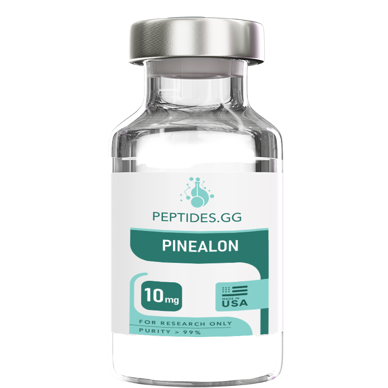
Buy Pinealon peptide for research applications. High-purity Pinealon research peptide available for laboratory studies and scientific investigation. Shop premium quality research-grade peptides manufactured in the USA with comprehensive Certificate of Analysis documentation.
Important: All products are intended as research chemicals only for laboratory and in vitro testing and experimentation. All product information is educational and not to be taken as medical advice. No products are for human or animal use.
Pinealon
$35.00
- Free Delivery on all orders over $200
- Earn 5% Store Credit with Every Order
- Same Day Shipping Before 1 PM PST
- 10% Discount for Cryptocurrency Payments
14-day money-back guarantee
If you are not satisfied with the product, simply return it and we will refund your money

Buy Pinealon peptide for research applications. High-purity Pinealon research peptide available for laboratory studies and scientific investigation. Shop premium quality research-grade peptides manufactured in the USA with comprehensive Certificate of Analysis documentation.
Important: All products are intended as research chemicals only for laboratory and in vitro testing and experimentation. All product information is educational and not to be taken as medical advice. No products are for human or animal use.
Research Overview
Pinealon serves as a valuable research tool for investigating neuroprotective mechanisms and brain tissue function in laboratory settings. As a synthetic tripeptide bioregulator originally derived from the pineal gland tissue of young animals, Pinealon represents a class of short peptides that demonstrate tissue-specific regulatory properties in experimental models. Research applications span neuroendocrine regulation, neuronal protection pathways, cellular senescence studies, and age-related neurological investigation.
Laboratory studies utilize Pinealon to examine brain-specific molecular interactions and their downstream biological effects on neuronal function. The pineal gland origin of this peptide provides unique research opportunities for investigating circadian rhythm regulation, melatonin production pathways, and neuroendocrine signaling mechanisms. Research protocols are designed to characterize peptide uptake into neural tissues, cellular responses in neuronal culture systems, and molecular pathway activation related to neuroprotection and cellular homeostasis.
The compound demonstrates specific biological activities in neurological tissue research models, making it valuable for diverse applications in neuroscience, gerontology, and cellular aging studies. Investigations examine mechanisms of neuroprotection, protein expression modulation in brain tissues, antioxidant pathway activation, and cellular stress response regulation in neural cells. Pinealon’s classification as a bioregulator peptide positions it among compounds studied for tissue-specific regulatory functions rather than receptor agonist or antagonist activities.
Research interest in Pinealon extends to its potential role in age-related neurological changes and cellular senescence processes. Studies investigate how short regulatory peptides might influence gene expression patterns in brain tissues, modulate protein synthesis in aging neurons, and affect cellular repair mechanisms. The peptide’s small size and specific amino acid composition allow for investigation of structure-activity relationships in bioregulator peptide research.
Molecular Characteristics
Complete Specifications:
- CAS Registry Number: NOT ASSIGNED
- Molecular Weight: 329.4 Da
- Molecular Formula: C14H23N5O6
- Amino Acid Sequence: Glu-Asp-Arg (EDR tripeptide)
- Peptide Classification: Synthetic tripeptide bioregulator
- Appearance: White to off-white lyophilized powder
- Solubility: Water, bacteriostatic water, phosphate buffered saline
- Tissue Specificity: Brain and pineal gland tissue research
The tripeptide molecular structure (Glu-Asp-Arg) contributes to specific biological activities investigated in neural tissue research settings. The sequence contains two acidic residues (glutamic acid and aspartic acid) and one basic residue (arginine), creating a net charge that influences cellular uptake, tissue distribution, and potential interactions with nuclear material in research models. Structural features of this short peptide allow for investigation of blood-brain barrier penetration, neuronal cell membrane interactions, and nuclear translocation mechanisms.
Structural Analysis:
The EDR tripeptide sequence exhibits specific physicochemical properties relevant to research applications. Glutamic acid (Glu) at position 1 provides a negatively charged carboxyl side chain at physiological pH, contributing to water solubility and potential electrostatic interactions with cellular components. Aspartic acid (Asp) at position 2 adds a second acidic residue with a shorter side chain than glutamate, influencing the peptide’s overall conformation and charge density. Arginine (Arg) at position 3 contributes a positively charged guanidinium group that participates in hydrogen bonding and electrostatic interactions with negatively charged cellular structures including nucleic acids and phospholipid membranes.
This charge distribution creates a zwitterionic peptide with both acidic and basic character, facilitating solubility across various pH ranges and potentially enabling cellular membrane translocation through multiple mechanisms. Research examines whether this specific amino acid arrangement allows for direct membrane penetration, receptor-mediated uptake, or transporter-facilitated cellular entry in neuronal tissue models. The small molecular weight (329.4 Da) positions Pinealon well below typical size limitations for blood-brain barrier passage, making it valuable for investigating peptide penetration into central nervous system tissues.
Conformational Properties:
Short tripeptides typically exhibit conformational flexibility in solution, allowing multiple structural configurations. Research investigates whether specific conformations of the EDR sequence contribute to biological activities or whether the peptide functions through its constituent amino acids following cellular uptake and potential hydrolysis. Circular dichroism spectroscopy and nuclear magnetic resonance studies could characterize solution conformations and stability profiles under various conditions relevant to experimental protocols.
Pharmacokinetic Profile in Research Models
Pinealon pharmacokinetic characterization in preclinical research provides important considerations for experimental design:
Absorption and Distribution:
- Demonstrates peptide uptake into brain tissues in experimental models
- Short peptide structure may facilitate cellular penetration mechanisms
- Research protocols investigate various administration routes for tissue distribution
- Bioavailability studies examine peptide stability and tissue-specific accumulation
Metabolic Stability:
- Tripeptide structure subject to peptidase degradation pathways
- Research examines peptide half-life in biological fluids and tissues
- Stability studies inform optimal experimental timing and dosing protocols
- Investigation of metabolite formation and biological activity of breakdown products
Tissue-Specific Effects:
- Research focuses on pineal gland and broader neural tissue distribution
- Studies examine peptide localization within cellular compartments
- Investigation of nuclear translocation and potential gene expression effects
- Duration of biological effects studied independently from plasma peptide levels
These pharmacokinetic characteristics inform research protocol design, particularly regarding timing of sample collection, dose-response relationships, and selection of appropriate experimental endpoints in neurological tissue studies.
Research Applications
Neuroprotection and Neuronal Survival Studies
Pinealon serves as a research tool for investigating neuroprotective mechanisms and neuronal cell survival pathways under various stress conditions. Laboratory studies examine the peptide’s effects on neuronal viability in cell culture models exposed to oxidative stress, excitotoxicity, or nutrient deprivation. Research protocols utilize primary neuronal cultures, neuronal cell lines (PC12, SH-SY5Y), and co-culture systems to assess protective effects against common neurotoxic insults.
Experimental approaches investigate Pinealon’s influence on apoptotic pathway modulation, examining caspase activation, mitochondrial membrane potential changes, and pro-apoptotic protein expression in stressed neuronal cells. Studies analyze antioxidant enzyme expression (SOD, catalase, glutathione peroxidase) and oxidative damage markers (lipid peroxidation, protein carbonylation, DNA oxidation) to characterize antioxidant mechanisms. Research examines calcium homeostasis regulation, investigating how the peptide might affect intracellular calcium levels and calcium-dependent signaling pathways in neural cells under excitotoxic conditions. These investigations provide insight into potential neuroprotective mechanisms at the cellular and molecular level.
Cellular Senescence and Aging Research
Research applications focus on Pinealon’s effects on cellular senescence markers and age-related changes in neuronal cells. Studies investigate senescence-associated beta-galactosidase activity, cell cycle arrest markers (p16, p21, p53), and senescence-associated secretory phenotype (SASP) factors in aging cell culture models. Research protocols examine telomere length, telomerase activity, and replicative capacity in cells exposed to the peptide over extended culture periods.
Investigation of age-related protein accumulation, examining effects on protein aggregation, proteasome activity, and autophagy pathway function in neuronal cells. Studies analyze expression of aging-associated genes and proteins, utilizing senescent cell models and comparing gene expression profiles with and without peptide exposure. Research examines mitochondrial function parameters including ATP production, oxygen consumption rates, and mitochondrial biogenesis markers in aging cell models. This research area explores bioregulator peptide effects on fundamental cellular aging processes and their potential modulation of age-related neurological changes in experimental models.
Gene Expression and Protein Synthesis Modulation
Pinealon research investigates potential effects on gene expression patterns in brain tissue models. Studies utilize transcriptomic approaches (RNA sequencing, microarray analysis) to examine global gene expression changes in neuronal cells or brain tissue samples exposed to the peptide. Research focuses on identification of specific gene pathways affected, examining neurotrophic factor expression, synaptic protein genes, antioxidant enzyme genes, and cell survival pathway genes.
Protein synthesis studies investigate the peptide’s effects on translation processes, examining both global protein synthesis rates and specific protein expression changes. Research protocols utilize Western blot analysis, immunohistochemistry, and proteomics approaches to characterize protein-level changes in experimental models. Investigation of nuclear translocation mechanisms examines whether the short tripeptide enters cell nuclei and potentially interacts with DNA or chromatin structures. Studies analyze immediate early gene expression, examining c-fos, c-jun, and other rapid-response genes to characterize cellular activation patterns. This research area addresses fundamental questions about bioregulator peptide mechanisms at the transcriptional and translational level.
Pineal Gland Function and Circadian Rhythm Research
Research applications specifically examine Pinealon’s effects on pineal gland tissue and circadian rhythm-related functions. Studies investigate melatonin production pathways, examining expression and activity of key enzymes (tryptophan hydroxylase, AANAT, HIOMT) involved in melatonin synthesis. Research utilizes pineal organ cultures, pinealocyte cell cultures, and tissue samples to assess effects on melatonin secretion patterns under various lighting conditions and circadian timing protocols.
Investigation of circadian clock gene expression (Per, Cry, Clock, Bmal1) in neural tissues and pineal cells examines potential chronobiological effects. Research protocols analyze circadian rhythm entrainment, phase shifting responses, and amplitude of circadian oscillations in cell culture and tissue models. Studies examine serotonin-to-melatonin conversion efficiency and regulation of adrenergic receptor expression in pinealocytes. Additional research investigates pineal gland morphology, cellular composition, and age-related changes in experimental models, providing tissue-specific characterization of bioregulator peptide effects on this neuroendocrine organ.
Neurotrophic Factor and Synaptic Plasticity Studies
Research examines Pinealon’s potential effects on neurotrophic factor expression and synaptic function in experimental models. Studies investigate brain-derived neurotrophic factor (BDNF), nerve growth factor (NGF), and other neurotrophins at mRNA and protein levels in neuronal cultures and brain tissue samples. Research protocols assess downstream signaling pathways including TrkB receptor activation, CREB phosphorylation, and ERK/MAPK pathway activation following peptide exposure.
Synaptic plasticity research utilizes hippocampal slice cultures, examining long-term potentiation (LTP), long-term depression (LTD), and synaptic protein expression (synaptophysin, PSD-95, synapsin). Studies analyze dendritic spine density and morphology using confocal microscopy and electrophysiological recordings to assess functional synaptic changes. Investigation of neurite outgrowth in neuronal culture models examines axon extension, branching patterns, and growth cone dynamics. Research explores acetylcholinesterase activity and cholinergic system function, examining effects on cognitive function-related pathways in experimental models. These investigations characterize potential neuroplasticity-related mechanisms of bioregulator peptides in neural tissue research.
Neuroinflammation and Glial Cell Research
Pinealon research applications include investigation of neuroinflammatory processes and glial cell function. Studies examine microglial activation states, analyzing morphological changes, pro-inflammatory cytokine production (IL-1beta, IL-6, TNF-alpha), and inflammatory mediator release (nitric oxide, prostaglandins) in microglial cell cultures and brain tissue models. Research protocols investigate NF-kappaB pathway activation, NLRP3 inflammasome assembly, and polarization of microglial phenotypes (M1 vs M2) in response to inflammatory stimuli with and without peptide exposure.
Astrocyte research examines glial fibrillary acidic protein (GFAP) expression, astrocyte reactivity, and astrocyte-neuron metabolic coupling. Studies investigate glutamate uptake capacity, examining excitatory amino acid transporter (EAAT) expression and function in astrocyte cultures. Research analyzes glial cell survival under stress conditions and the role of astrocytic support in neuronal survival experiments. Blood-brain barrier modeling studies utilize endothelial cell cultures and co-culture systems to examine tight junction protein expression and barrier integrity. This research area addresses the complex cellular interactions in neural tissue and potential immunomodulatory effects of bioregulator peptides.
Mitochondrial Function and Bioenergetics Research
Research investigates Pinealon’s effects on mitochondrial function in neuronal and neural tissue models. Studies examine mitochondrial membrane potential using fluorescent probes (TMRM, JC-1), assessing mitochondrial polarization status under basal and stressed conditions. Research protocols analyze cellular bioenergetics using Seahorse technology or Clark electrode measurements, examining oxygen consumption rates, ATP production, and respiratory chain complex activities.
Mitochondrial biogenesis studies investigate PGC-1alpha expression, mitochondrial DNA content, and expression of mitochondrial proteins in cells exposed to the peptide. Research examines mitochondrial dynamics, analyzing fission and fusion processes through assessment of Drp1, Mfn1/2, and OPA1 protein expression and localization. Studies investigate reactive oxygen species (ROS) production at the mitochondrial level, examining superoxide generation and antioxidant capacity. Research analyzes mitochondrial calcium handling, examining calcium uniporter function and mitochondrial calcium buffering capacity. These investigations characterize cellular energy metabolism and mitochondrial health parameters relevant to neurological function and neuroprotection mechanisms.
Cognitive Function and Memory-Related Pathway Research
Research applications examine molecular pathways associated with cognitive function and memory formation in experimental models. Studies investigate synaptic transmission efficiency, examining neurotransmitter release, receptor expression (NMDA, AMPA glutamate receptors), and postsynaptic signaling mechanisms. Research protocols utilize hippocampal tissue and cell models to examine molecular correlates of learning and memory, including calcium/calmodulin-dependent protein kinase II (CaMKII) activity and protein kinase A/C signaling.
Investigation of acetylcholine system function examines choline acetyltransferase expression, acetylcholinesterase activity, and cholinergic receptor expression in neural tissue models. Research analyzes immediate early gene expression patterns associated with synaptic activity and memory consolidation processes. Studies examine adult neurogenesis markers in hippocampal cultures or tissue samples, investigating effects on neural progenitor cell proliferation, differentiation, and survival. Molecular pathway analysis focuses on CREB-dependent transcription, examining phosphorylated CREB levels and CREB-target gene expression related to memory formation. This research provides insight into cognitive function-related molecular mechanisms in experimental neurological research models.
DNA Repair and Genomic Stability Research
Research applications investigate Pinealon’s potential effects on DNA repair mechanisms and genomic stability in neuronal cells. Studies examine expression and activity of DNA repair enzymes including base excision repair (BER) pathway components (OGG1, APE1, DNA polymerase beta), nucleotide excision repair proteins, and mismatch repair factors in neuronal culture models exposed to oxidative stress or genotoxic agents. Research protocols analyze DNA damage markers including 8-oxo-deoxyguanosine formation, single-strand breaks, and double-strand breaks using comet assay, immunofluorescence techniques, and Western blot analysis.
Investigation of cell cycle checkpoint regulation examines p53 expression and phosphorylation, checkpoint kinase activation (Chk1, Chk2), and cell cycle arrest mechanisms in response to DNA damage with and without peptide exposure. Research analyzes histone modifications associated with DNA repair including histone H2AX phosphorylation (gamma-H2AX foci formation) and chromatin remodeling factors. Studies examine telomere integrity and telomere-associated DNA damage in aging neuronal models, investigating whether bioregulator peptides influence telomere maintenance mechanisms. This research area addresses fundamental questions about peptide effects on genomic stability and DNA damage response pathways in neural cells, particularly relevant to age-related neurological research where accumulated DNA damage contributes to cellular dysfunction. Experimental protocols utilize both basal conditions and DNA damage-inducing treatments to characterize protective or reparative mechanisms.
Endoplasmic Reticulum Stress and Protein Folding Research
Research examines Pinealon’s effects on endoplasmic reticulum (ER) stress pathways and protein homeostasis in neuronal cells. Studies investigate unfolded protein response (UPR) activation, examining expression and phosphorylation of UPR sensors including PERK, IRE1alpha, and ATF6 in neuronal models exposed to ER stress inducers (tunicamycin, thapsigargin, brefeldin A). Research protocols analyze downstream UPR signaling including eIF2alpha phosphorylation, CHOP expression, spliced XBP1 production, and ATF4 transcription factor activity using quantitative PCR, Western blotting, and luciferase reporter assays.
Investigation of ER-associated degradation (ERAD) examines proteasomal degradation of misfolded proteins and expression of ERAD components in stressed neuronal cells. Research analyzes ER chaperone expression including BiP/GRP78, GRP94, protein disulfide isomerase, and calnexin, investigating whether peptide exposure modulates ER chaperone capacity and protein folding assistance. Studies examine calcium homeostasis at the ER level, analyzing ER calcium stores, calcium release patterns, and ER-mitochondria calcium transfer in neuronal models. Research investigates ER morphology using electron microscopy and fluorescent ER markers, examining ER network organization and potential ER fragmentation under stress conditions. This research area is particularly relevant to neurodegenerative disease models where protein misfolding and ER stress contribute to pathological processes. Experimental approaches characterize whether bioregulator peptides can modulate ER stress responses and enhance neuronal proteostasis capacity under challenging conditions.
Laboratory Handling and Storage Protocols
Lyophilized Powder Storage:
- Store at -20°C to -80°C in original sealed vial
- Protect from light exposure and moisture
- Desiccated storage environment recommended
- Stability data available for 12+ months at -20°C
Reconstitution Guidelines:
- Reconstitute with sterile water, bacteriostatic water (0.9% benzyl alcohol), or appropriate buffer
- Add solvent slowly down vial side to minimize foaming and peptide aggregation
- Gentle swirling motion recommended (avoid vigorous shaking which may denature peptide)
- Allow complete dissolution before use (typically 30-60 seconds for tripeptide)
- Final pH should be 7.0-8.0 for optimal stability in aqueous solution
- Typical reconstitution concentration: 1-10 mg/mL depending on experimental requirements
- Filter sterilization (0.22 micron) recommended if preparing stock solutions for cell culture work
- Document reconstitution date, concentration, and storage conditions for laboratory records
Reconstituted Solution Storage:
- Short-term: 4°C for up to 7 days
- Long-term: -20°C in single-use aliquots
- Avoid repeated freeze-thaw cycles (maximum 2-3 cycles)
Quality Assurance and Analytical Testing
Each batch undergoes comprehensive analytical characterization:
Purity Analysis:
- High-Performance Liquid Chromatography (HPLC): ≥98% purity
- Reversed-phase HPLC with UV detection
- Multiple peak integration for accurate determination
Structural Verification:
- Mass Spectrometry (ESI-MS): Confirms molecular weight
- Amino acid analysis: Verifies composition
- Peptide content determination: Quantifies actual content
Contaminant Testing:
- Bacterial endotoxin: <5 EU/mg (LAL method)
- Heavy metals: Below detection limits
- Residual solvents: Within acceptable limits
- Water content: Karl Fischer titration (<8%)
Documentation:
- Certificate of Analysis provided with each batch
- Third-party verification available upon request
- Stability data for recommended storage conditions
- Batch-specific results traceable by lot number
Research Considerations
Researchers should consider several factors when designing experiments with Pinealon:
1. Experimental Design: Determine appropriate concentrations based on research objectives
2. Model Selection: Choose suitable experimental systems aligned with research questions
3. Controls: Include appropriate vehicle controls and comparative compounds
4. Temporal Factors: Consider pharmacokinetic characteristics in timing measurements
5. Reproducibility: Follow consistent protocols for reliable results
Mechanism investigation requires careful experimental design to isolate specific effects and pathways. Multiple approaches may be needed to fully characterize biological activities.
Experimental Protocol Examples
Neuroprotection Assay in Primary Neuronal Cultures:
Primary cortical neurons isolated from embryonic day 18 rat embryos are cultured in neurobasal medium supplemented with B27, glutamine, and antibiotics. After 7 days in vitro when neuronal maturation is established, cultures are pretreated with Pinealon at concentrations ranging from 0.1 to 100 micromolar for 24 hours. Oxidative stress is then induced using hydrogen peroxide (50-200 micromolar) or glutamate excitotoxicity (100 micromolar) for 3-6 hours. Neuronal viability is assessed using MTT assay, lactate dehydrogenase release measurement, or live/dead cell staining with calcein-AM and ethidium homodimer. Additional endpoints include caspase-3/7 activity measurement for apoptosis assessment, measurement of intracellular reactive oxygen species using DCF-DA fluorescence, and Western blot analysis of apoptotic markers (cleaved caspase-3, Bax, Bcl-2) at 6-24 hours post-injury. This protocol allows quantification of neuroprotective effects and investigation of underlying mechanisms through multiple complementary measurements.
Gene Expression Analysis in Neuronal Cell Lines:
SH-SY5Y human neuroblastoma cells or PC12 rat pheochromocytoma cells are plated at 5×10^5 cells per well in 6-well plates and allowed to attach overnight. Cells are then treated with Pinealon (10 micromolar) or vehicle control for designated time periods (3, 6, 12, 24 hours) to examine temporal gene expression patterns. Following treatment, cells are harvested in TRIzol reagent for RNA extraction, and total RNA is isolated using standard chloroform extraction and isopropanol precipitation protocols. RNA quality is verified by spectrophotometry (A260/A280 ratio) and gel electrophoresis before proceeding to reverse transcription using random hexamer primers or oligo-dT. Quantitative real-time PCR examines expression of genes of interest including neurotrophic factors (BDNF, NGF, GDNF), antioxidant enzymes (SOD1, SOD2, catalase, GPX1), anti-apoptotic factors (Bcl-2, Bcl-xL), and immediate early genes (c-fos, Arc, Egr1). Results are normalized to reference genes (GAPDH, beta-actin, HPRT) and analyzed using delta-delta Ct method. For broader transcriptome analysis, RNA sequencing can be performed to identify global gene expression changes induced by peptide treatment.
Mitochondrial Function Assessment Protocol:
Neuronal cells (primary neurons or neuronal cell lines) are seeded in XF96 microplates at optimized density (20,000-40,000 cells per well) and cultured for 24 hours before treatment with Pinealon at various concentrations. Following 24-48 hour treatment periods, cellular bioenergetics are analyzed using Seahorse XF96 Extracellular Flux Analyzer measuring oxygen consumption rate and extracellular acidification rate. The mitochondrial stress test protocol sequentially injects oligomycin (ATP synthase inhibitor, 1 micromolar), FCCP (mitochondrial uncoupler, 0.5-2 micromolar titrated for optimal response), and rotenone/antimycin A (complex I and III inhibitors, 0.5 micromolar each). From the resulting oxygen consumption rate measurements, key parameters are calculated including basal respiration, ATP production-linked respiration, proton leak, maximal respiration capacity, spare respiratory capacity, and non-mitochondrial respiration. Parallel experiments assess mitochondrial membrane potential using TMRM or JC-1 fluorescent indicators, mitochondrial reactive oxygen species production using MitoSOX Red, and ATP levels using luminescence-based assays. This comprehensive approach characterizes multiple aspects of mitochondrial function and bioenergetics.
Immunofluorescence Microscopy for Cellular Localization:
Neuronal cells grown on poly-L-lysine coated glass coverslips in 24-well plates are treated with Pinealon (10-50 micromolar) for various time periods (30 minutes to 24 hours) to examine peptide effects on cellular structures and protein localization. Following treatment, cells are fixed with 4% paraformaldehyde for 15 minutes, permeabilized with 0.1% Triton X-100 for 10 minutes, and blocked with 5% normal goat serum for 1 hour at room temperature. Primary antibodies targeting proteins of interest (synaptic markers, cytoskeletal proteins, nuclear proteins, mitochondrial markers) are applied overnight at 4 degrees Celsius at appropriate dilutions. After washing, fluorophore-conjugated secondary antibodies are applied for 1-2 hours at room temperature, followed by nuclear counterstaining with DAPI. Coverslips are mounted on slides using anti-fade mounting medium and examined using confocal microscopy with appropriate laser lines. Quantitative analysis includes measurement of fluorescence intensity, protein colocalization analysis using Pearson’s or Manders’ coefficients, automated cell counting for positive/negative cells, and morphological measurements (neurite length, dendritic spine density). This approach visualizes cellular and subcellular effects of peptide treatment.
Synaptic Plasticity Investigation in Hippocampal Slices:
Acute hippocampal slices (350-400 micrometers thick) are prepared from young adult rodents using a vibratome in ice-cold cutting solution. Slices are transferred to artificial cerebrospinal fluid and allowed to recover for at least 1 hour at room temperature before experimentation. For electrophysiological recordings, slices are perfused with oxygenated artificial cerebrospinal fluid at 30-32 degrees Celsius, and recording electrodes are placed in the CA1 stratum radiatum while stimulating electrodes activate Schaffer collateral pathways. Following 20 minutes of stable baseline recording at low frequency stimulation, Pinealon is applied to the perfusion solution at designated concentrations. Long-term potentiation is induced using high-frequency stimulation protocols (100 Hz for 1 second, repeated 2-3 times) or theta-burst stimulation, and field excitatory postsynaptic potentials are recorded for 60-120 minutes post-induction. Slice viability is confirmed by paired-pulse facilitation testing. Additional experiments examine effects on basal synaptic transmission through input-output curve analysis, presynaptic function through paired-pulse ratio measurements, and synaptic protein expression in slice lysates through Western blotting. This ex vivo approach maintains tissue architecture while allowing controlled experimental manipulations and direct electrophysiological assessment of synaptic function.
Compliance and Safety Information
Regulatory Status:
Pinealon is provided as a research chemical for in-vitro laboratory studies and preclinical research only. Not approved by FDA for human therapeutic use.
Intended Use:
- In-vitro cell culture studies
- In-vivo preclinical research
- Laboratory mechanism investigation
- Academic and institutional research
NOT Intended For:
- Human consumption or administration
- Therapeutic treatment or diagnosis
- Dietary supplementation
- Veterinary therapeutic applications
Safety Protocols:
- Use appropriate personal protective equipment
- Handle in well-ventilated areas
- Follow institutional biosafety guidelines
- Dispose of waste per local regulations
- Consult MSDS for additional safety information
—
 25% OFF $400+ • 35% OFF $800+ ALL WEEKEND


